- Research article
- Open Access
- Published:
Expression patterns of ciliopathy genesARL3andCEP120reveal roles in multisystem development
BMC Developmental Biologyvolume20, Article number:26(2020)
Abstract
Background
Joubert syndrome and related disorders (JSRD) and Jeune syndrome are multisystem ciliopathy disorders with overlapping phenotypes. There are a growing number of genetic causes for these rare syndromes, including the recently described genesARL3andCEP120.
Methods
We sought to explore the developmental expression patterns ofARL3andCEP120in humans to gain additional understanding of these genetic conditions. We used an RNA in situ detection technique called RNAscope to characteriseARL3andCEP120expression patterns in human embryos and foetuses in collaboration with the MRC-Wellcome Trust Human Developmental Biology Resource.
Results
BothARL3andCEP120are expressed in early human brain development, including the cerebellum and in the developing retina and kidney, consistent with the clinical phenotypes seen with pathogenic variants in these genes.
Conclusions
This study provides insights into the potential pathogenesis of JSRD by uncovering the spatial expression of two JSRD-causative genes during normal human development.
Background
Joubert syndrome and related disorders (JSRD) are a group of autosomal inherited ciliopathies that are characterised as a cerebello-retinal-renal phenotype, and have an incidence rate of 1:80,000–100,000 live births [1,2,3]. The hallmark brain phenotype is a “molar tooth sign” shown on axial brain MRI, caused by cerebellar vermis hypoplasia and other mid and hindbrain malformations [4]. These defects often cause symptoms of hypotonia, ataxia and intellectual disability in patients [5]. The retinal and renal phenotypes associated with JSRD have a lower incidence rate and vary in severity. Renal disorders occur in ~ 25% of patients, often presenting as corticomedullary cysts, interstitial fibrosis, or tubulointerstitial kidney disease [5]. The renal component is progressive and can lead to end-stage renal disease [6]. Ocular phenotypes of retinitis dystrophy, retinitis pigmentosa, oculomotor apraxia, and ptosis are common in patients, and as with the renal aspects of JSRD are often progressive in nature [7].
Currently, there are more than 35 genes that are known to cause JSRD (https://www.omim.org/phenotypicSeries/PS213300). The syndrome is caused by defects of the primary cilia, which are found on most mammalian cells [8]. Primary cilia act as a cellular antenna to transduce extracellular signals such as mechanical flow, chemical stimulation, and key signalling pathways (including Hedgehog, Wnt, and PDGF) into the cell [9,10,11,12,13]. Due to the multi-organ involvement, varying phenotypes, and multitude of genes known to cause JSRD there is great heterogeneity within the syndrome and overlap with closely related ciliopathies including Bardet-Biedl syndrome (https://omim.org/phenotypicSeries/PS209900) and Jeune syndrome (https://omim.org/phenotypicSeries/PS208500) [14]. Recently discovered genetic causes of JSRD includeARL3[15] andCEP120[16,17]; the fact that their encoded proteins have such divergent roles within the primary cilium demonstrates the complexity underlying this group of related disorders.
ADP-ribosylation factor-like 3 (ARL3), a RAS superfamily member, is a low molecular weight GTP-binding protein [18] that cycles between inactive GDP-bound and active GTP-bound states to release cargo from their carriers in the cilium [19]. ARL3 interacts with its Guanine Exchange Factor ARL13B in the cilium [20] and GTPase Activating Protein RP2 at the basal body of the cell [21,22].Arl3knockout studies in mice demonstrate a multi-organ ciliopathy phenotype, including kidney cysts, liver fibrosis and retinal disease with photoreceptor cell degeneration [23,24,25,26]. Recently, two families affected by JSRD have been identified, presenting with ciliopathy phenotypes [15]. The underlying genetic cause was shown to be missense mutations inARL3, which affect an amino acid residue involved in the interaction between ARL3 and ARL13B [15].
Centrosomal protein of 120 kDa (CEP120) is a centrosomal protein involved in centriole biogenesis, including centriole duplication, assembly [27,28], elongation [29,30,31] and maturation [28]. CEP120 also interacts with other centrosomal proteins including CPAP [29,30], SPICE1 [29], Talpid3 [28,31] and C2CD3 [31]. CEP120 was found to be expressed ubiquitously in murine embryonic tissues such as the brain, kidney and lungs. Additionally, Cep120 was observed to be highly expressed in embryonic mouse brain compared to postnatal or adult mouse brain [32]. Inactivation ofCEP120in the mouse central nervous system results in hydrocephalous and cerebellar hypoplasia [28].CEP120突变已被证明导致JSRD和另一幅作品《年轻syndromes [16,17], and overlapping ciliopathy phenotypes such as tectocerebellar dysraphia with occipital encephalocele (TCDOE), Meckel syndrome (MKS) and oro-facial-digital (OFD) syndromes [17].
The developmental expression patterns ofARL3andCEP120in humans is not known. In order to explore this, we used an RNA in situ detection technique called RNAscope to compare and contrast the developmental spatial expression of these new and divergent causes of JSRD. We successfully characterisedARL3andCEP120expression patterns in human embryos and foetuses in collaboration with the MRC-Wellcome Trust Human Developmental Biology Resource (HDBR). This study provides insights into the potential pathogenesis of JSRD by uncovering the expression pattern of two JSRD-causative genes during normal human development.
Methods
RNAscope studies
Characterisation ofARL3andCEP120expression patterns was performed in human embryonic tissue using samples obtained from the MRC-Wellcome HDBR. Formalin fixed paraffin embedded sections of human embryonic and foetal tissue were prepared using 10% neutral buffered formalin and fixed for 32 h at room temperature. Samples were then prepared for the RNAscope assay, a RNA in situ detection platform for detection of target RNA within intact cells, as per manufacturers’ instructions [33,34]. An RNAscope 2.5 Assay RED was employed with 20 paired probes across nucleotides 169–1570 (NM_004311.3) and 115–1133 (NM_001166226.1) for detection ofARL3andCEP120, respectively and counterstained with Methyl Green.
Whole human embryo sections of 8 post-conception weeks (PCW), (equivalent to Carnegie Stage 23) were analysed, along with hindbrain (14PCW and 19PCW), eye (14PCW), kidney and adrenal gland (14PCW and 18PCW). A negative RNAscope 2.5 HD Assay Red control (dapB, a bacterial gene which is not expressed in human tissues) was performed (Supplementary Figure1). In addition, the RNAscope 2.5 HD Assay RED was performed with a positive control (KI67, a cell proliferation marker). KI67 is a nuclear protein commonly used as a proliferation marker, which is expressed in cycling cells and is associated with cellular proliferation. It is encoded by the gene marker of proliferation Ki-67,MKI67. ARL3andCEP120human expression patterns were analysed using the HDBR image server (Leica Biosystems).
Clinical phenotypes and sequence analysis
Reported clinical phenotypes associated withARL3andCEP120mutations were reviewed within OMIM (https://omim.org/). Putative Arl3 and Cep120 orthologues were identified using BLASTP, with human ARL3 (isoform a, NP_004302.1) and CEP120 (NP_694955.2) transcripts as the query sequences within NCBI (https://www.ncbi.nlm.nih.gov/). Additional databases including Flybasehttps://flybase.org/), Wormbase (https://www.wormbase.org/) and Phytozome (https://phytozome.jgi.doe.gov/) were also queried using BLAST.
Results
Clinical phenotypes of ARL3 and CEP120 patients
Biallelic mutations in bothARL3andCEP120mutations are rare causes of ciliopathy syndromes. A comparison of the known phenotypes associated withARL3andCEP120mutations is shown in Table1. This overview reveals that mutations inCEP120are at present associated with severe phenotypes including MKS but also that single heterozygous changes inARL3are sufficient to cause retinal-limited phenotypes.ARL3is highly conserved, with homologs present inC. elegans,C. reinhardtiiandD. melanogasterwhereasCEP120appears not to have homologs within these lower organisms (Supplementary Table1). Known protein localisation within the cell of both ARL3 within the ciliary axoneme, and CEP120 in the centrosomes, are consistent with their role in ciliopathy syndromes (Supplementary Table2).
ARL3 and CEP120 are expressed in early human brain development
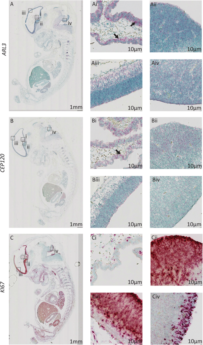
In 8PCW human brain tissue, the expression ofARL3andCEP120is remarkably similar. There is expression of both genes in the choroid plexus (Fig.1Ai and 1Bi), which appears to favour the luminal facing surface of the tissue, especially forARL3. The cell proliferation markerKI67does not share this same expression pattern in the choroid plexus (Fig.1 Ci). This specific expression pattern ofARL3andCEP120in luminal-facing cells is continued throughout the developing brain where both genes exhibit expression throughout the ventricular zone of the ganglionic eminences, cortical wall, and the hindbrain including the rhombic lip (Fig.1Aii-Aiv and 1Bii-Biv). There is specific expression ofARL3andCEP120in the layer of cells forming the apical surface in each tissue, facing into the ventricular space. Expression ofKI67is seen throughout these tissues (Fig.1Cii-Civ),与特定的表达式在顶端layer consistent with this being the major site of cell division (cells in G2/M1 phase of cell cycle) in the ventricular zone [37].
Expression pattern ofARL3andCEP120in the human brain during early development.Sagittal sections of 8PCW-stage human embryos stained using RNAscope to show expression ofARL3(a) (red),CEP120(b) (red) andKI67(c) (red), counterstained with Methyl Green.AiandBiARL3andCEP120are expressed within cells of the choroid plexus (arrow). (Ci)KI67expression is minimal in the choroid plexus.Aii-Aivand (Bii-Biv) Expression ofARL3andCEP120is seen in the ventricular radial glia progenitor cells including the ventricular zone of the ganglionic eminences (AiiandBii), cerebral cortex (AiiiandBiii), and rhombic lip (AivandBiv).Cii-CivExpression ofKI67is seen in the ventricular zone of the ganglionic eminences, cerebral cortex, and hindbrain
Expression of ARL3 and CEP120 is maintained in the developing cerebellum
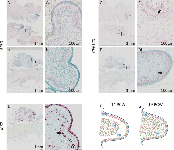
In the human cerebellum at 14PCW there is expression ofARL3andCEP120. Both genes have strong expression in the external and internal granule cell layer (EGL and IGL) the developing cerebellum (Fig.2Ai and Ci). Expression in the EGL and IGL is seen at 19PCW forARL3(Fig2Bi) however,CEP120expression is predominantly localised in the EGL and the molecular layer (ML) of the cerebellum at 19PCW (Fig.2Di). Strong expression ofKI67is seen throughout the EGL in particular, but also the IGL at 19PCW, indicating the tissue is proliferative (Fig.2Ei).ARL3andCEP120are therefore widely expressed in the cerebellum during development, with specific expression ofCEP120in the ML which is predominantly occupied by the dendritic trees of Purkinje cells and the interacting parallel fibres of granule cells. As dendrites and axons contain low levels of mRNA, it is likely thatCEP120expression is predominantly located in the sparse population of interneurons found in the molecular layer [38] or in immature granule cells migrating from EGL to IGL [39] suggesting a role forCEP120in these cell types [38].
Expression ofARL3andCEP120in the developing human hindbrain. Sagittal sections of 14PCW (aandc) and 19PCW (b,dande) human brain stained using RNAscope to showARL3expression (aandb) (red),CEP120(candd) (red) andKI67(e) (red), countered stained with Methyl Green.AiandCiExpression ofARL3andCEP120is evident in the cerebellum at 14PCW.banddARL3andCEP120expression in the cerebellum at 19PCW.BiCerebellar expression ofARL3.Di小脑分子层(箭头)的表达CEP120.eandEiHindbrainKI67expression at 18PCW shows tissue is proliferative.fandgSchematic diagrams of developing cerebellum at (f) 14PCW and (g) 19PCW. Expression ofARL3is shown in red andCEP120in green. EGL, external granule layer; IGL, internal granule layer; ML, molecular layer
Expression of ARL3 and CEP120 in the developing eye
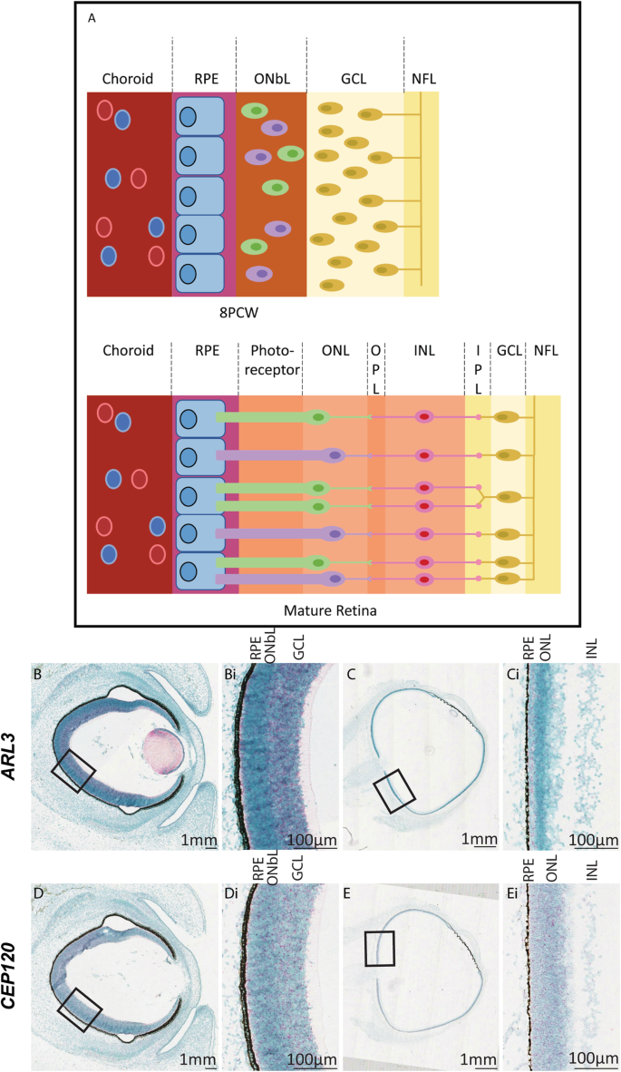
The human retina can be divided into nine layers based upon the cell types that occupy each layer (Fig.3a), with the retinal pigment epithelial (RPE) and photoreceptor layers at the outermost part of the eye [39]. At 8PCW, the retinal layers are not well defined with only a ganglion cell layer separated from a layer of mostly immature neuroblasts with a few photoreceptor cells by a thin inner plexiform layer [40]. At this stageARL3andCEP120show expression throughout the developing retina, with high expression within the retinal ganglion cells and the photoreceptor layer (Fig.3Bi and Di). At 14PCW, the retinal layers are maturing [39] which is reflected in the expression pattern of bothARL3andCEP120.Clear expression of both genes is still seen in all layers of the retina, although to a lesser extent in the plexiform and nerve fibre layers due to reduced cell density in these areas (Fig.3 Ci and Ei).
Expression ofARL3andCEP120in the developing human retina.aSchematic diagram of the development of the layers of the retina from 8PCW to the mature form (adapted from [40]). At 8PCW, not all of the layers are present in the retina. The ONbL is a transitionary layer containing retinal progenitor cells that will develop into various cell types such as photoreceptors, amacrine and bipolar cells; separating into the ONL, OPL, INL, and IPL (the IPL is sometimes visible at 8PCW). The GCL is thicker at 8PCW due to the migration of cells. The mature retina can be divided into layers. The RPE is at the very back of the eye and assists in the removal of waste products from the photoreceptor cells, which transduce light. The ONL, OPL, INL and IPL layers house intermediary cell bodies and dendrites that interact with ganglion cells in the GCL to convey the signal through the optic nerve, formed in the NFL, to the brain (reviewed in [41]).bHuman sections of developing eye at 8PCW (bandd) and 14PCW (cande) stained using RNA Scope to show ARL3 expression (bandc) (red) and CEP120 (dande) (red) counterstained with Methyl Green (BiandDi). There is a gradient of ARL3 (Bi) and CEP120 (Di) expression in the retina at 8PCW across multiple retinal layers including the ONbL. At 14 PCW, ARL3 (Ci) and CEP120 (Ei) expression is localised across multiple layers including the photoreceptor cell layer, just below the RPE layer (arrows). GCL, ganglion cell layer; INL, inner nuclear layer; IPL, inner plexiform layer; NFL, nerve fibre layer; ONL, outer nuclear layer; OPL, outer plexiform layer; ONbL, outer neuroblastic layer; RPE, retinal pigment epithelium
Expression of ARL3 and CEP120 in the developing dorsal root ganglia
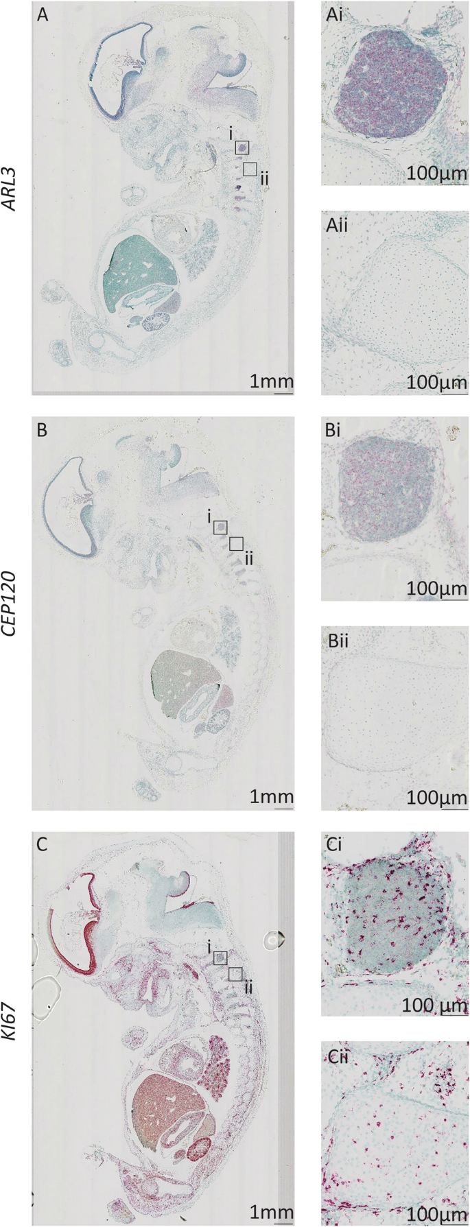
The dorsal root ganglia are formed by migrating neural crest cells and contain most of the body’s sensory neurones [42,43]. BothARL3andCEP120show expression in cells of the dorsal root ganglia, which are post-mitotic primary sensory neurons (Fig.4Ai-Aii and Bi-Bii). There is strong expression ofKI67within limited number of cells in the dorsal root ganglia, presumably non-neuronal (Fig.4 Ci-Cii).
Expression ofARL3andCEP120in the developing human dorsal root ganglia Sagittal sections of 8PCW human embryos stained using RNAscope to show expression ofARL3(a) (red),CEP120(b) (red) andKI67(c) (red) counterstained with Methyl Green.ARL3andCEP120expression is shown within the dorasl root ganglia (AiandBirespectively), whereas surrounding tissue has low level expression of these genes (AiiandBii).KI67expression is seen in the dorsal root ganglia (Ci) and surrounding tissues (Cii)
Expression of ARL3 and CEP120 in the developing kidney
In the developing human kidney at 8PCW, where there is strong renal cortical staining ofKI67indicating cell proliferation (Supplementary Figure2), there is expression ofARL3细胞在发展中皮质肾单位;this expression appears to be specifically oriented to the lumen of the structures (Fig.5Ai). This expression pattern is maintained at 14PCW (Fig.5Bi) and 18PCW (Fig.5 Ci). Expression ofCEP120is also seen in developing nephrons at 8PCW, however there is also expression in the renal cortex (Fig.5Di). This expression pattern ofCEP120is maintained at 14PCW (Fig.5Ei) and 18PCW, although overall expression appears to have decreased at this time point (Fig.5Fi).
Expression ofARL3andCEP120in the developing human kidney. Sagittal sections of human kidney at 8PCW (aandd), 14PCW (bande), and 18PCW (candf) stained using RNAscope to showARL3expression (a,b,c) (red) andCEP120(d,e,f) (red) and counterstained with Methyl Green.ARL3andCEP120expression at 8PCW (AiandDi) is seen in the developing kidney cortex.ARL3andCEP120expression in the kidney cortex remain the same at 14PCW (BiandEirespectively). Ci and Fi shows persistentARL3and reducedCEP120 kidney cortex expression at 18PCW
Expression of ARL3 and CEP120 in other major organs
In the developing human heart, lung and gut at 8PCW, there is very low levels of expression ofARL3andCEP120(Supplementary Figure3). Expression ofARL3andCEP120is seen around the developing alveoli and at low levels in the developing bowel epithelia. The remaining organs of the developing embryo did not reveal prominent expression patterns.
Discussion
Mutations inARL3andCEP120are rare and relatively new causes of JSRD and other related ciliopathies. Human protein atlas data suggests that tissue expression of ARL3 protein is widely expressed, with highest expression scores seen in cerebellum and lowest in heart and skeletal muscle (https://www.proteinatlas.org/ENSG00000138175-ARL3/tissue). RNA expression is high in cerebral cortex, cerebellum, retina and kidney consistent with its known phenotypes. CEP120 protein expression is not annotated within the human protein atlas, whereas RNA is strongly expressed in the cerebellum (https://www.proteinatlas.org/ENSG00000168944-CEP120/tissue). We aimed to define expression ofARL3andCEP120during human development using the HDBR tissue bank employing a relatively new in situ hybridisation assay called RNAscope for the detection of target RNA within intact cells. We usedKI67as a positive control although we recognise that expression ofKI67is not homogeneous throughout each tissue. Our data provide an insight into the developmental expression ofARL3andCEP120. We show that both of these genes are expressed in key tissues (including retina, cerebellum and kidney) during development. This expression pattern fits with the multisystem disease phenotypes seen in patients withARL3andCEP120mutations (Table1). A similar approach, using the valuable HDBR tissue bank has been performed, using in situ hybridisation for studying the expression ofARL13B[44],another cause of Joubert syndrome. HereARL13B发现在腋下和基底阶段CS16 plate of the myelencephalon, the mesencephalon and the metencephalon. At CS19ARL13Bwas seen in the ventricular layer of the diencephalon and myelencephalon, the tegmentum of the pons and the cerebellar rhombic lips as well as the dorsal root ganglia. This pattern of expression is remarkably similar to theCEP120andARL3data described here.
Expression of bothARL3andCEP120was minimal in developing cardiac, lung and gut tissues, consistent with lack of known phenotypes affecting these organ systems (Supplementary Figure3).ARL3andCEP120encode proteins that are expressed in the primary cilia and basal body respectively (Supplementary Table2) and pathogenic variants result in similar and overlapping phenotypes, including the cerebello-retinal-renal syndrome JSRD (Table1). The number of patients with pathogenic variants in eitherARL3orCEP120remains small, allowing a limited comparison of phenotypes, although skeletal manifestations (in particular short ribs/asphyxiating thoracic dystrophy phenotypes) seen in patients withCEP120mutations have not been documented in patients withARL3mutations.
There were notable differences in evolutionary conservation betweenARL3andCEP120(Supplementary Table1). The ARL3 human protein shares greater than 90% identity with its two orthologous sequences (there is genomic duplication ofarl3) inDanio renio(zebrafish), a well-studied model species in vertebrates. In contrast, CEP120 human protein only shares 57% identity with its single orthologous sequence found in zebrafish. Moreover, human ARL3 protein shares > 60% identity with its orthologues found inDrosophila melanogaster,Caenorhabditis elegansandChlamydomonas reinhardtii. CEP120 is conserved in some vertebrate organisms but orthologues were not readily identified in invertebrates. There is a putativeCEP120orthologue, UNI2, found inChlamydomonas reinhardtii, but this has not as yet been confirmed as a functional ortholog [27,44]. ARL3 is described in diverse eukaryotic organisms such asLeishmania donovani[45] andCaenorhabditis elegans[46] [47] where it has a functional role in the cilium/flagella. Despite these differences in evolutionary conservation, our results show thatARL3andCEP120have similar expression patterns during human development, specifically in the eye and dorsal root ganglia as well as during early brain development. Both genes are expressed throughout the retina during development, with expression in the RPE and photoreceptor layers, suggesting a role for both genes during retinal development. This is further supported by the numerous retinal phenotypes associated with mutations inARL3[15,16,17]. Similarly, the specific expression ofARL3andCEP120in the dorsal root ganglia hints at a role for both genes in primary sensory neurone differentiation. A recurring pattern was the expression of both mRNAs on the luminal facing surface of the cerebral tissue (seen in the choroid plexus and ventricular zones of the cerebral cortex ganglionic eminences, and hindbrain) which could suggest a sensory role for the gene products of both the genes within the cilium of the ventricular lining of the brain.
Expression ofARL3andCEP120changes during development notably in the cerebellum and kidney.ARL3andCEP120are expressed throughout the cerebellum at 14PCW however, at 19PCW, ARL3 expression was predominantly in the IGL whereasCEP120was expressed in the EGL and ML of the cerebellum. This could imply thatARL3andCEP120are expressed in different cell populations of the cerebellum,ARL3in both immature and mature granule cells, andCEP120in immature, migratory granule cells and ML interneurones [48]. It has been previously reported in mouse studies thatCep120is required for proliferation of cerebellar neural progenitor cells [28] and is required for correct development of the embryo. Taken with these results, it suggests thatCEP120expression is required for correct development of the cerebellum in humans.
Expression ofARL3andCEP120also differed in the developing kidney. The results showed thatARL3was specifically expressed in cells of the nephrons whereasCEP120was expressed in the nephrons as well as within cells in the developing renal cortex. This difference in expression could imply thatARL3has a more sensory/signalling function in luminal structures of the kidney, whereasCEP120has a more universal role in all cells as it is expressed more ubiquitously throughout the tissue.
The differences in gene expression may reflect the divergent functions of ARL3 and CEP120 proteins (Supplementary Table2). As ARL3 is a trafficking protein involved in ciliary signalling [19,49], it may only be expressed in actively signalling cells during certain points in development such as nephron progenitors and cells in the IGCL. In contrast, CEP120 is involved in building the centriole, and therefore cilium, [27,29] and so will be expressed more widely within tissues, especially those with ciliated epithelia [50,51].
In conclusion, we establish in human embryonic tissue expression patterns ofARL3andCEP120during development and provide insights into the wide phenotypic spectrum of mutations affectingARL3andCEP120in humans. These studies will allow further investigations into tissue-specific mechanistic roles ofARL3andCEP120in human health and disease.
Availability of data and materials
All data generated or analysed during this study are included in this published article and its supplementary information files. The datasets used and/or analysed during the current study are available from the corresponding author on reasonable request.
Abbreviations
- EGL:
-
External granule cell layer
- GCL:
-
Ganglion cell layer
- HDBR:
-
Human Developmental Biology Resource
- IGL:
-
Internal granule cell layer
- INL:
-
Inner nuclear layer
- IPL:
-
Inner plexiform layer
- JATD:
-
Jeune asphyxiating thoracic dystrophy
- JSRD:
-
Joubert syndrome and related disorders
- MKS:
-
Meckel syndrome
- ML:
-
Molecular layer
- NFL:
-
Nerve fibre layer
- OFD:
-
Oro-facial-digital
- ONbL:
-
Outer neuroblastic layer
- ONL:
-
Outer nuclear layer
- OPL:
-
Outer plexiform layer
- PCW:
-
Post-conception weeks
- RPE:
-
Retinal pigment epithelium
- TCDOE:
-
Tectocerebellar dysraphia with occipital encephalocele
References
Malicdan MC, Vilboux T, Stephen J, Maglic D, Mian L, Konzman D, et al. Mutations in human homologue of chicken talpid3 gene (KIAA0586) cause a hybrid ciliopathy with overlapping features of Jeune and Joubert syndromes. J Med Genet. 2015;52(12):830–9.
Travaglini L, Brancati F, Silhavy J, Iannicelli M, Nickerson E, Elkhartoufi N, et al. Phenotypic spectrum and prevalence of INPP5E mutations in Joubert syndrome and related disorders. Eur J Human Genet. 2013;21(10):1074–8.
Valente EM, Brancati F, Dallapiccola B. Genotypes and phenotypes of Joubert syndrome and related disorders. Eur J Med Genet. 2008;51(1):1–23.
Doherty D. Joubert syndrome: insights into brain development, cilium biology, and complex disease. Semin Pediatr Neurol. 2009;16(3):143–54.
Paprocka J, Jamroz E. Joubert syndrome and related disorders. Neurol Neurochir Pol. 2012;46(4):379–83.
Srivastava S, Ramsbottom SA, Molinari E, Alkanderi S, Filby A, White K, et al. A human patient-derived cellular model of Joubert syndrome reveals ciliary defects which can be rescued with targeted therapies. Hum Mol Genet. 2017;26(23):4657–67.
Wang SF, Kowal TJ, Ning K, Koo EB, Wu AY, Mahajan VB, et al. Review of Ocular Manifestations of Joubert Syndrome. Genes. 2018;9:12.
Reiter JF, Leroux MR. Genes and molecular pathways underpinning ciliopathies. Nat Rev Mol Cell Biol. 2017;18(9):533–47.
Berbari NF, O'Connor AK, Haycraft CJ, Yoder BK. The primary cilium as a complex signaling center. Curr Biol. 2009;19(13):R526–35.
Malicki JJ, Johnson CA. The cilium: cellular antenna and central processing unit. Trends Cell Biol. 2017;27(2):126–40.
Christensen ST, Pedersen LB, Schneider L, Satir P. Sensory cilia and integration of signal transduction in human health and disease. Traffic (Copenhagen, Denmark). 2007;8(2):97–109.
Hildebrandt F,奥托·e·纤毛中心体:联合国ifying pathogenic concept for cystic kidney disease? Nat Rev Genet. 2005;6(12):928–40.
Wheway G, Parry DA, Johnson CA. The role of primary cilia in the development and disease of the retina. Organogenesis. 2014;10(1):69–85.
Fansa EK, Wittinghofer A. Sorting of lipidated cargo by the Arl2/Arl3 system. Small GTPases. 2016;7(4):222–30.
Alkanderi S, Molinari E, Shaheen R, Elmaghloob Y, Stephen LA, Sammut V, et al. ARL3 mutations cause Joubert syndrome by disrupting Ciliary protein composition. Am J Hum Genet. 2018;103(4):612–20.
Shaheen R, Schmidts M, Faqeih E, Hashem A, Lausch E, Holder I, et al. A founder CEP120 mutation in Jeune asphyxiating thoracic dystrophy expands the role of centriolar proteins in skeletal ciliopathies. Hum Mol Genet. 2015;24(5):1410–9.
Roosing S, Romani M, Isrie M, Rosti RO, Micalizzi A, Musaev D, et al. Mutations in CEP120 cause Joubert syndrome as well as complex ciliopathy phenotypes. J Med Genet. 2016;53(9):608–15.
Kahn RA, Der CJ, Bokoch GM. The ras superfamily of GTP-binding proteins: guidelines on nomenclature. FASEB J. 1992;6(8):2512–3.
Gotthardt K, Lokaj M, Koerner C, Falk N, Giessl A, Wittinghofer A. A G-protein activation cascade from Arl13B to Arl3 and implications for ciliary targeting of lipidated proteins. eLife. 2015;4.
Blacque OE, Perens EA, Boroevich KA, Inglis PN, Li C, Warner A, et al. Functional genomics of the cilium, a sensory organelle. Curr Biol. 2005;15(10):935–41.
Veltel S, Gasper R, Eisenacher E, Wittinghofer A. The retinitis pigmentosa 2 gene product is a GTPase-activating protein for Arf-like 3. Nat Struct Mol Biol. 2008;15(4):373–80.
Evans RJ, Schwarz N, Nagel-Wolfrum K, Wolfrum U, Hardcastle AJ, Cheetham ME. The retinitis pigmentosa protein RP2 links pericentriolar vesicle transport between the Golgi and the primary cilium. Hum Mol Genet. 2010;19(7):1358–67.
Grayson C, Bartolini F, Chapple JP, Willison KR, Bhamidipati A, Lewis SA, et al. Localization in the human retina of the X-linked retinitis pigmentosa protein RP2, its homologue cofactor C and the RP2 interacting protein Arl3. Hum Mol Genet. 2002;11(24):3065–74.
Schwarz N, Lane A, Jovanovic K, Parfitt DA, Aguila M, Thompson CL, et al. Arl3 and RP2 regulate the trafficking of ciliary tip kinesins. Hum Mol Genet. 2017;26(13):2480–92.
Hanke-Gogokhia C, Wu Z, Gerstner CD, Frederick JM, Zhang H, Baehr W. Arf-like protein 3 (ARL3) regulates protein trafficking and Ciliogenesis in mouse photoreceptors. J Biol Chem. 2016;291(13):7142–55.
Schrick JJ, Vogel P, Abuin A, Hampton B, Rice DS. ADP-ribosylation factor-like 3 is involved in kidney and photoreceptor development. Am J Pathol. 2006;168(4):1288–98.
Mahjoub MR, Xie Z, Stearns T. Cep120 is asymmetrically localized to the daughter centriole and is essential for centriole assembly. J Cell Biol. 2010;191(2):331–46.
Wu C, Yang M, Li J, Wang C, Cao T, Tao K, et al. Talpid3-binding centrosomal protein Cep120 is required for centriole duplication and proliferation of cerebellar granule neuron progenitors. PLoS One. 2014;9(9):e107943.
Comartin D, Gupta GD, Fussner E, Coyaud E, Hasegan M, Archinti M, et al. CEP120 and SPICE1 cooperate with CPAP in centriole elongation. Curr Biol. 2013;23(14):1360–6.
Lin YN, Wu CT, Lin YC, Hsu WB, Tang CJ, Chang CW, et al. CEP120 interacts with CPAP and positively regulates centriole elongation. J Cell Biol. 2013;202(2):211–9.
Tsai JJ, Hsu WB, Liu JH, Chang CW, Tang TK. CEP120 interacts with C2CD3 and Talpid3 and is required for centriole appendage assembly and ciliogenesis. Sci Rep. 2019;9(1):6037.
Xie Z, Moy LY, Sanada K, Zhou Y, Buchman JJ, Tsai LH. Cep120 and TACCs control interkinetic nuclear migration and the neural progenitor pool. Neuron. 2007;56(1):79–93.
Wang H, Wang MX, Su N, Wang LC, Wu X, Bui S, et al. RNAscope for in situ detection of transcriptionally active human papillomavirus in head and neck squamous cell carcinoma. Journal of visualized experiments : JoVE. 2014;85.
Wang F, Flanagan J, Su N, Wang LC, Bui S, Nielson A, et al. RNAscope: a novel in situ RNA analysis platform for formalin-fixed, paraffin-embedded tissues. J Mol Diagnostics. 2012;14(1):22–9.
Holtan JP, Teigen K, Aukrust I, Bragadottir R, Houge G. Dominant ARL3-related retinitis pigmentosa. Ophthalmic Genet. 2019;40(2):124–8.
Strom SP, Clark MJ, Martinez A, Garcia S, Abelazeem AA, Matynia A, et al. De novo occurrence of a variant in ARL3 and apparent autosomal dominant transmission of retinitis Pigmentosa. PLoS One. 2016;11(3):e0150944.
Taverna E, Huttner WB. Neural progenitor nuclei IN motion. Neuron. 2010;67(6):906–14.
Hatten ME, Heintz N. Mechanisms of neural patterning and specification in the developing cerebellum. Annu Rev Neurosci. 1995;18:385–408.
Chalupa LM, Gunhan E. Development of on and off retinal pathways and retinogeniculate projections. Prog Retin Eye Res. 2004;23(1):31–51.
Hendrickson A. Development of Retinal Layers in Prenatal Human Retina. Am J Ophthalmology. 2016;161:29–35 e1.
Hoon M, Okawa H, Della Santina L, Wong ROL. Functional architecture of the retina: development and disease. Prog Retin Eye Res. 2014;42:44–84.
Krames ES. The role of the dorsal root ganglion in the development of neuropathic pain. Pain Med (Malden, Mass). 2014;15(10):1669–85.
Kalcheim C, Barde YA, Thoenen H, Le Douarin NM. In vivo effect of brain-derived neurotrophic factor on the survival of developing dorsal root ganglion cells. EMBO J. 1987;6(10):2871–3.
Piasecki BP, Silflow CD. The UNI1 and UNI2 genes function in the transition of triplet to doublet microtubules between the centriole and cilium in Chlamydomonas. Mol Biol Cell. 2009;20(1):368–78.
Cuvillier A, Redon F, Antoine JC, Chardin P, DeVos T, Merlin G. LdARL-3A, a Leishmania promastigote-specific ADP-ribosylation factor-like protein, is essential for flagellum integrity. J Cell Sci. 2000;113(Pt 11):2065–74.
Li Y, Wei Q, Zhang Y, Ling K, Hu J. The small GTPases ARL-13 and ARL-3 coordinate intraflagellar transport and ciliogenesis. J Cell Biol. 2010;189(6):1039–51.
Sharma A, Gerard SF, Olieric N, Steinmetz MO. Cep120 promotes microtubule formation through a unique tubulin binding C2 domain. J Struct Biol. 2018;203(1):62–70.
Sotelo C. Molecular layer interneurons of the cerebellum: developmental and morphological aspects. Cerebellum (London, England). 2015;14(5):534–56.
Ismail SA, Chen YX, Rusinova A, Chandra A, Bierbaum M, Gremer L, et al. Arl2-GTP and Arl3-GTP regulate a GDI-like transport system for farnesylated cargo. Nat Chem Biol. 2011;7(12):942–9.
Tucker RW, Pardee AB, Fujiwara K. Centriole ciliation is related to quiescence and DNA synthesis in 3T3 cells. Cell. 1979;17(3):527–35.
Pugacheva EN, Jablonski SA, Hartman TR, Henske EP, Golemis EA. HEF1-dependent Aurora a activation induces disassembly of the primary cilium. Cell. 2007;129(7):1351–63.
Acknowledgements
Schematic diagrams were created usingBiorender.com
Funding
LP is funded by the Medical Research Council Discovery Medicine North Training Partnership. MB-G is funded by Kidney Research UK (ST_001_20171120) and Northern Counties Kidney Research Fund. EM is funded by Kidney Research UK (Paed_RP_20180925). SAR is Kidney Research UK post-doctoral fellow (PDF_003_20151124). CGM and JAS are funded by Kidney Research UK and Northern Counties Kidney Research Fund. The funding bodies had no role in the design of the study and collection, analysis, and interpretation of data nor in the writing of the manuscript.
Author information
Authors and Affiliations
Contributions
LP and MB-G analysed and interpreted the data regarding the RNA expression studies. GJC, LAD, SAR and CGM performed data analysis and contributed in writing the manuscript. JAS conceived the project. All authors read and approved the final manuscript.
Corresponding author
Ethics declarations
Ethics approval and consent to participate
This study was conducted with full ethical approval. For human embryonic and foetal tissue samples, the samples were collected with appropriate written maternal consents and ethical approval by the Newcastle and North Tyneside 1 Research Ethics Committee, UK.
Consent for publication
Not applicable.
Competing interests
The authors declare that they have no competing interests.
Additional information
Publisher’s Note
Springer Nature remains neutral with regard to jurisdictional claims in published maps and institutional affiliations.
鲍威尔L和Barroso-Gil M是共同第一作者
Supplementary Information
12861_2020_231_MOESM1_ESM.pdf
Additional file 1.
Rights and permissions
Open Access本文是在Creative Commons许可Attribution 4.0 International License, which permits use, sharing, adaptation, distribution and reproduction in any medium or format, as long as you give appropriate credit to the original author(s) and the source, provide a link to the Creative Commons licence, and indicate if changes were made. The images or other third party material in this article are included in the article's Creative Commons licence, unless indicated otherwise in a credit line to the material. If material is not included in the article's Creative Commons licence and your intended use is not permitted by statutory regulation or exceeds the permitted use, you will need to obtain permission directly from the copyright holder. To view a copy of this licence, visithttp://creativecommons.org/licenses/by/4.0/. The Creative Commons Public Domain Dedication waiver (http://creativecommons.org/publicdomain/zero/1.0/) applies to the data made available in this article, unless otherwise stated in a credit line to the data.
About this article
Cite this article
Powell, L., Barroso-Gil, M., Clowry, G.J.et al.Expression patterns of ciliopathy genesARL3andCEP120reveal roles in multisystem development.BMC Dev Biol20, 26 (2020). https://doi.org/10.1186/s12861-020-00231-3
Received:
Accepted:
Published:
DOI:https://doi.org/10.1186/s12861-020-00231-3
Keywords
- CEP120
- ARL3
- Foetus
- Development
- Retina
- Kidney
- Brain
- RNAscope


